
By | Medically Reviewed by Christopher W. Boelter, MD | Last updated 2026-07-14
A Kidney Stone Can Hurt Badly, but Pain Is Only Part of the Story
Most people picture a kidney stone as an episode of unbearable pain that starts without warning. That does happen. A man may be working, driving, or sleeping when a sharp pain suddenly grips one side of his back. He cannot find a comfortable position. The pain eases for a few minutes, then returns. Nausea, sweating, or an urgent need to urinate may follow.
But not every stone behaves that way. Some remain in the kidney and cause no symptoms. Others produce an ache that is easy to mistake for a pulled muscle. A stone can also interfere with urine drainage without creating the worst pain a patient has ever felt.
Kidney stones in men are solid deposits formed from substances concentrated in urine. Their effect depends on where they are, whether urine can drain around them, whether infection is present, and how well the kidneys are functioning. Stone size matters, but it is not the only factor.
The intensity of pain does not reliably tell us whether a kidney stone is dangerous. Fever, chills, reduced urine output, uncontrolled vomiting, a solitary functioning kidney, and signs of infection can be more important than the number a patient gives the pain on a scale from 1 to 10.
This guide is written for the man who wants a practical answer: Does this sound like a stone, can I wait, and what will happen when I seek care? It also explains why flank pain should not be diagnosed at home. Appendicitis, gallbladder disease, aortic disease, kidney infection, spinal problems, hernia, and testicular conditions can overlap with stone symptoms.
Why the Pain Moves From the Back Toward the Groin
A stone may sit quietly inside the kidney. Symptoms often begin when it moves into the ureter, the narrow muscular tube that carries urine from the kidney to the bladder. The kidney continues to make urine above the stone. Pressure can build behind the obstruction while the ureter contracts in an effort to move the stone downward.
This wave-like pain is called renal colic. It often begins in the flank, between the lower ribs and the hip. As the stone moves down the ureter, the discomfort may shift toward the side of the abdomen, the lower abdomen, the groin, or the testicle.
Patients often ask what kidney stone pain feels like. The honest answer is that the pattern varies, but several features are common:
- Pain begins in the side or back, often on one side
- The intensity rises and falls in waves
- The patient may feel restless and unable to find a comfortable position
- Pain may travel toward the lower abdomen, groin, or testicle
- Nausea, vomiting, sweating, urinary urgency, or burning may occur
| Stone location | Common pain pattern | Other possible clues |
|---|---|---|
| Inside the kidney | No pain or a dull ache below the ribs | Blood found on a urine test, repeated infection, or an incidental imaging finding |
| Upper ureter | Sharp pain in the flank or back | Nausea, vomiting, sweating, and restlessness |
| Middle ureter | Pain moving toward the side or lower abdomen | Waves of pain and blood in the urine |
| Lower ureter | Lower abdominal, groin, or referred testicular pain | Urgency, frequency, burning, or pressure near the bladder |
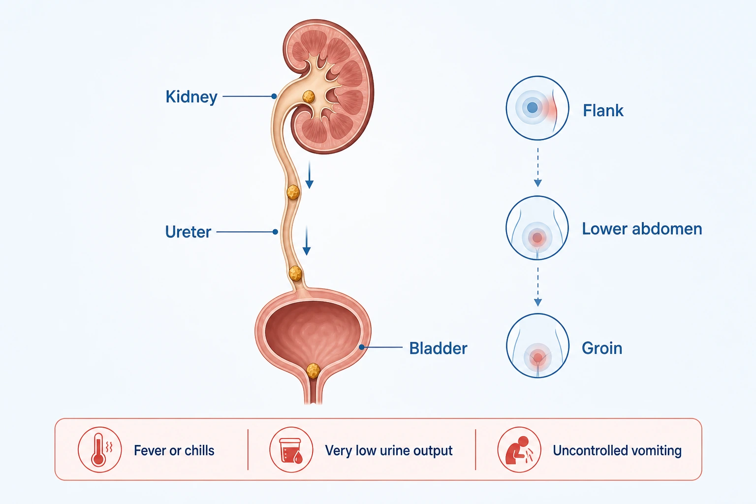
A ureteral stone near the bladder may feel more like a urinary infection than classic back pain. The patient may urinate frequently, feel a constant urge to go, or experience burning. Pain referred to the testicle can occur, but sudden severe one-sided scrotal pain should never be blamed on a kidney stone without evaluation. The pattern described in testicular pain that requires urgent care can indicate torsion or another time-sensitive problem.
When Waiting at Home Is Not Safe
The combination clinicians worry about most is urinary obstruction with infection. If a stone blocks drainage and bacteria are present above it, pressure and infection can build inside the kidney. Antibiotics may be necessary, but they may not be enough if infected urine cannot drain.
An obstructed and infected kidney is a urologic emergency that may require immediate drainage as well as antibiotics. Drainage is usually achieved with a ureteral stent or a nephrostomy tube. Definitive stone removal is often postponed until the infection is controlled and the patient is stable.
The question of when a kidney stone is an emergency should be answered from the whole situation. Seek urgent or emergency medical care if possible stone symptoms occur with:
- Fever, shaking chills, confusion, faintness, or rapidly worsening weakness
- Very low urine output or no urine output
- Inability to urinate despite a painful or full bladder
- Repeated vomiting or inability to keep fluids down
- Pain that cannot be controlled with appropriately used medication
- One functioning kidney, a transplanted kidney, or significant chronic kidney disease
- Heavy visible bleeding, clots, or increasing difficulty passing urine
- Severe pain when a stone has not previously been confirmed
Not being able to empty a full bladder can represent acute urinary retention in men. That is different from a stone blocking one ureter, although both problems can cause pain and reduced urination.
A familiar history of stones does not make every new episode routine. If the pain changes location, fever appears, urine output falls, or the patient looks and feels seriously ill, the condition needs to be reassessed.
What Blood and Other Urine Changes Can Mean
Blood in urine is common with stones because a moving stone can irritate the lining of the kidney or ureter. The urine may look pink, red, brown, or tea-colored. Sometimes the blood is microscopic and appears only during laboratory testing.
Blood does not prove that a stone is present, and a urine sample without blood does not rule one out. Infection, kidney disease, prostate conditions, trauma, medicines, and urinary tract cancers can also cause bleeding. For that reason, the appropriate evaluation of hematuria depends on age, smoking history, previous bleeding, infection findings, and other individual risks.
A patient should also report cloudy urine, unpleasant odor, burning, frequency, urgency, or reduced output. Cloudiness and odor alone do not diagnose infection. Fever, urinary pain, flank pain, and systemic illness are more concerning and may lead to urinalysis, culture, blood testing, and urgent imaging.
Stones and poor urinary drainage can contribute to recurrent urinary tract infections in men. When infections return, treating each episode without looking for an obstructing stone or another structural cause may leave the underlying problem unresolved.
What the Tests Actually Tell the Urologist
The right tests for kidney stones depend on the patient, the urgency of the situation, previous imaging, kidney function, and whether another diagnosis is possible. There is no single test that answers every question.
Urinalysis and Urine Culture
Urinalysis may show red blood cells, white blood cells, crystals, acidity, and other clues. Crystals do not automatically mean that a stone is blocking the ureter. White blood cells can occur with irritation as well as infection. A urine culture helps determine whether bacteria are growing and which antibiotics may be effective.
Blood Tests
Blood testing may assess kidney function, electrolytes, blood cell counts, calcium, uric acid, and signs of systemic infection. The exact tests depend on the presentation. A man who is feverish and unwell needs a different evaluation from someone whose painless stone was found incidentally.
Ultrasound
Ultrasound uses no ionizing radiation. It can identify some stones and show swelling of the kidney collecting system when drainage is impaired. It may miss certain ureteral stones or provide less precise information about their size and position.
Noncontrast Computed Tomography
Noncontrast computed tomography can detect most urinary stones, locate them, measure them, and show the degree of obstruction. It can also reveal some alternative explanations for acute pain. Radiation exposure still matters, especially in younger men who have repeated stone episodes, so scanning should be selected thoughtfully rather than repeated automatically.
Follow-Up Imaging
Pain relief does not always confirm that the stone has passed. Follow-up imaging may be used to determine whether the stone moved, whether obstruction resolved, or whether treatment was successful. The timing and type of imaging depend on the original findings and the patient’s progress.
A broader explanation of urinary tract stone diagnosis and treatment covers stones in the kidneys, ureters, bladder, and urethra. The location matters because it changes both the symptoms and the available treatment options.
Can a Stone Pass on Its Own?
Some stones pass without a procedure. Others do not. Size and location are important, but they are not the entire decision. The urologist also considers obstruction, infection, kidney function, pain control, nausea, anatomy, previous procedures, and whether the patient can return promptly if symptoms worsen.
Observation may be reasonable when:
- There is no evidence of infection
- Pain and nausea can be controlled
- Kidney function is acceptable
- The patient can drink and is not becoming dehydrated
- No high-risk kidney or urinary anatomy is present
- Follow-up and repeat testing can be completed safely
A patient may be asked to strain the urine and save the stone for analysis. If pain disappears but the stone is not seen, follow-up may still be needed. A blocked kidney can occasionally become less painful even though the obstruction remains.
Alpha-blockers may be discussed as medical expulsive therapy for selected ureteral stones. They are not appropriate for every patient or every stone, and this use may be off-label. Current European guidance reports the clearest possible benefit in selected distal ureteral stones larger than 5 mm. A patient should not borrow tamsulosin, change a dose, or begin treatment solely on the basis of an online article.
When a Procedure Is the Safer Choice
The choice between observation and a procedure depends on stone location, size, obstruction, infection, kidney function, symptom control, and the safety of follow-up. There is no responsible recommendation based only on the diameter printed in an imaging report.
A procedure may be recommended when the stone is unlikely to pass, pain remains uncontrolled, vomiting continues, obstruction threatens kidney function, infection is present, or observation has not worked.
Common options include:
- Ureteral stent placement to restore urine drainage
- Percutaneous nephrostomy drainage through the back into the kidney
- Ureteroscopy to see, fragment, and remove a stone
- Shock wave lithotripsy to break selected stones into smaller pieces
- Percutaneous nephrolithotomy for selected large or complex kidney stones
Emergency drainage and final stone removal are not always performed together. If a blocked kidney is infected, the first job is to drain the kidney and treat the infection. Definitive removal usually follows after the patient has stabilized.
Preventing Another Stone Starts With Knowing What Formed
“Drink more water” is useful advice, but it is not a complete prevention plan. Fluid needs vary with climate, physical activity, heart and kidney function, urinary symptoms, and the type of stone. A patient who has been told to restrict fluids should not ignore that instruction.
The best answer to how to prevent kidney stones from returning begins with stone analysis whenever a stone is passed or removed. Men with recurrent stones, unusual stone types, a strong family history, a solitary kidney, or other high-risk features may also need blood tests and one or more 24-hour urine collections.
Depending on the results, prevention may involve:
- Drinking enough fluid to produce an appropriate daily urine volume
- Reducing excessive sodium intake
- Maintaining an appropriate amount of calcium from food
- Adjusting oxalate, purine, or animal protein intake when indicated
- Treating urinary infection, gout, bowel disease, or another contributing condition
- Using potassium citrate, a thiazide-type medicine, allopurinol, or another preventive treatment when laboratory findings support it
Eliminating calcium from the diet without medical advice can be a mistake. For some calcium oxalate stone formers, an appropriate amount of dietary calcium helps bind oxalate in the digestive tract. Prevention should match the stone and the patient, not a generic list of forbidden foods.
What to Bring to a Urology Appointment
Kidney stone symptoms are easier to interpret when the timeline is clear. Write down where the pain began, where it moved, whether it came in waves, and whether fever, vomiting, urinary changes, or testicular discomfort occurred. Bring previous imaging reports, urine cultures, stone analysis, procedure notes, and a complete medicine and supplement list.
Useful questions include:
- Where is the stone, and is it blocking urine flow?
- Is there evidence of infection or reduced kidney function?
- Is it reasonable to wait for the stone to pass?
- How will we confirm that the obstruction has resolved?
- Which symptoms mean I should seek emergency care?
- Do I need stone analysis or a metabolic evaluation?
Patients who need a urology appointment can contact Adult & Pediatric Urology. Fever, inability to urinate, very low urine output, uncontrolled vomiting, or rapidly worsening illness should be directed to urgent or emergency care rather than a routine appointment request.
Medical Disclaimer
This article provides general medical education and does not diagnose a kidney stone or replace individualized care. Seek urgent medical attention for fever with urinary pain, inability to urinate, very low urine output, uncontrolled pain or vomiting, severe weakness, confusion, or rapidly worsening symptoms.
Frequently Asked Questions
What Does Kidney Stone Pain Usually Feel Like?
It often begins as sharp pain in the flank or back below the ribs and may move toward the lower abdomen, groin, or testicle. The pain can rise and fall in waves. Nausea, sweating, restlessness, urinary urgency, burning, or blood in urine may occur, but symptoms alone cannot confirm the diagnosis.
Can a Kidney Stone Cause Blood in Urine Without Severe Pain?
Yes. A stone may cause microscopic or visible blood with mild discomfort or no pain. Because infection, kidney disease, prostate conditions, medicines, trauma, and urinary tract cancers can also cause bleeding, visible or persistent blood should be evaluated.
When Is a Kidney Stone an Emergency?
Urgent assessment is needed when possible stone symptoms occur with fever, chills, confusion, very low urine output, inability to urinate, uncontrolled pain, repeated vomiting, severe dehydration, one functioning kidney, or rapidly worsening illness. Obstruction combined with infection is especially dangerous.
Which Tests Are Used to Diagnose a Kidney Stone?
Evaluation may include urinalysis, urine culture, blood tests, ultrasound, and noncontrast computed tomography. Computed tomography can locate most stones and show obstruction. Ultrasound avoids radiation and can identify collecting-system swelling and some stones. The appropriate test depends on urgency and individual factors.
Can Kidney Stones Be Prevented From Coming Back?
Risk can often be reduced after the stone type and contributing factors are identified. Prevention may include individualized fluid intake, sodium reduction, appropriate dietary calcium, treatment of related conditions, and selected medicines based on blood or 24-hour urine results.